Brain Tumor Extraction
Medical image processing is the most challenging and emerging field now a days. Processing of MRI images is one of the part of this field. This project describes the proposed strategy to detect & extraction of brain tumour from patient’s MRI scan images of the brain. This method incorporates with some noise removal functions, segmentation and morphological operations which are the basic concepts of image processing. Detection and extraction of tumour from MRI scan images of the brain is done by using MATLAB software.
METHODOLOGY
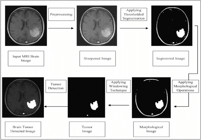
This project describes the strategy to detect and extract brain tumor from patient’s MRI scan images. First it takes the name and age of a person and then MRI brain image is used for tumor detection process. It includes pre-processing, segmentation, morphological operation, watershed segmentation and calculation of the tumor area and determination of the tumor location.
In this, a brain tumour MRI image is applied to pre-processing and after that tumour is extracted by morphological and watershed segmentation processes. The medical image segmentation has difficulties in segmenting complex structure with uneven shape, size, and properties. In such condition it is better to use unsupervised methods such as edge detection algorithm. For accurate diagnosis of tumor patients, appropriate segmentation method is required to be used for MRI images to carry out an improved diagnosis and treatment. The brain tumor detection is a great help for the physicians and a boon for the medical imaging and industries working on the production of MRI imaging.
FUTURE SCOPE
In near future, a database can be created for different patients having different types of brain tumours and locate them. Tumour growth can be analysed by plotting graph which can be obtained by studying sequential images of tumour affect. Possible extension of the presented work could use more features. It would be beneficial to connect the system to cloud storage of patient‟s information in hospital. This application can be extended to accessibility and usability through mobile phones. If this application is developed to analyse all types of MRI scans of same patient and result of all scans are integrated, it can suggest appropriate treatment and medication as well.